
At the imaging department of the hospital in Ísafjörður, general X-rays, CT scans and also electrocardiograms are carried out. In Patreksfjörður, general X-ray examinations are carried out. There must always be a medical reason for X-ray examinations and they must be carried out in an approved manner, so that radiation is as low as possible. Then the risk associated with the radiation is justified and it is guaranteed that the benefit to the patient is greater than the risk.
Patient radiation doses can vary greatly during the same X-ray examination, both between patients and between individual X-ray departments. However, the goal is the same everywhere; to keep radiation doses as low as possible.
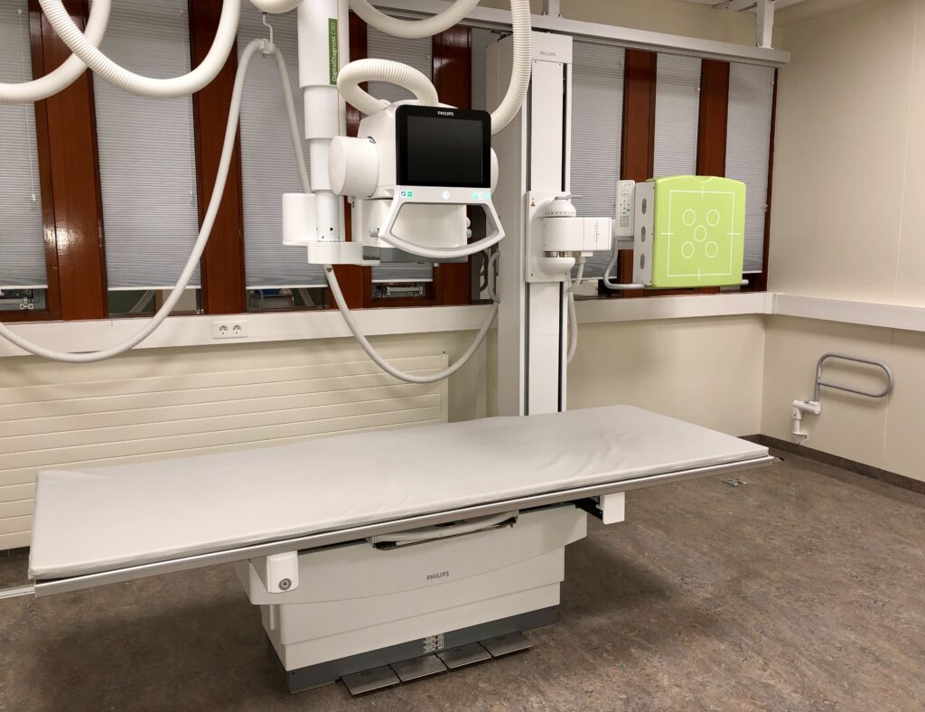
General X-ray examinations
An X-ray is used to take pictures of the body's bones, abdomen or lungs. Usually 2-4 pictures are taken, but the number of pictures depends on the research area.
Preparation
Clothing and jewelry that can obscure the area of the body to be photographed must be removed.
Execution
It is important to follow the radiologist's instructions, for example regarding breathing. Depending on the area to be imaged, you will need to sit, stand or lie on the exam bench. A radiologist adjusts you and steps aside to take an image, but the reason radiologists take images every day is that the amount of radiation in general x-rays is not dangerous. X-ray examinations take approx. 10-20 minutes.
Radiation protection is used as needed, but the most important protection lies in the precise and correct work practices of radiologists.
results
All images are sent to Domus Medica for reading. Radiologists analyze the images and send a response to the doctor who requested the examination, who is responsible for informing the patient about the results.
The reading usually takes 1-2 days, but you can stop by the reception and make an appointment with a doctor a few days after the test to access the results.
Children undergoing X-ray examination
Children do not feel it when an X-ray is taken, but it is important that the child is still during it. Parents are welcome to stay with their children and often need to help them stay completely still. If a parent is with their child, they must wear a lead apron during imaging to protect themselves from unnecessary radiation.
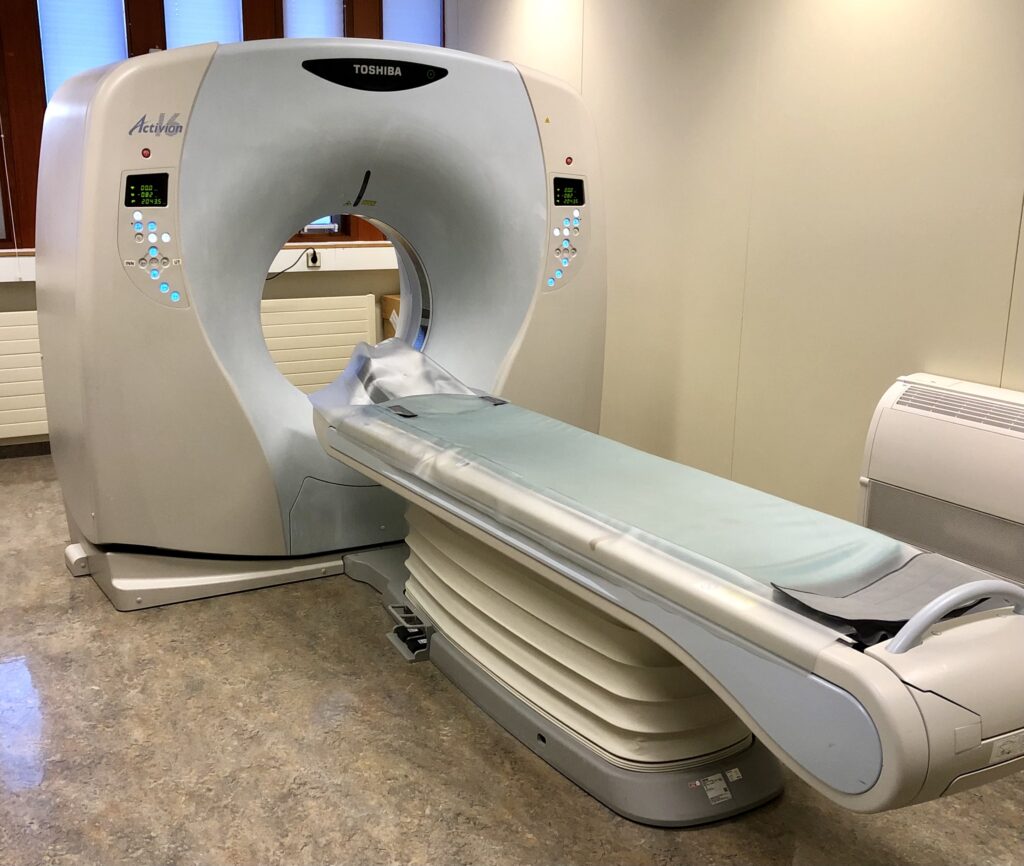
Computed tomography studies
An X-ray is used to obtain cross-sectional images of organs, organ systems or certain body parts. With a CT scan, you can get detailed information about the condition of organs and blood vessels and their position, as well as detect fractures in the bone that cannot be seen on a general X-ray.
Preparation
CT scan preparation depends on the area to be imaged and the reason for the study. Clothing must be removed if there is metal or metal threads in clothing, jewelry and other accessories that can obscure the image area or give off image defects.
Execution
During the examination, you have to lie on an examination bench that moves slowly back and forth into the CT scanner. The device most closely resembles a large doughnut, so there is little chance of feeling trapped. You are set up for the examination, so it is important to lie completely still afterwards. Sometimes an iodine contrast agent must be given intravenously and a catheter is then installed. A CT scan usually takes about 10-20 minutes.
Iodine contrast material is given intravenously and is used to distinguish between organs, assess their condition and make blood vessels visible on a picture. It is normal to feel heat in the body flowing from the throat down to the bladder, a feeling like urination is taking place and often a bad taste in the mouth. These reactions are normal and pass quickly.
Intolerance or allergic reaction can occur during the administration of iodine contrast material. If you have a known allergy to the contrast material, inform the doctor requesting the examination, the reception desk or the radiologist before the examination. It is important to notify in advance so that preparations can be started in time or another research method can be chosen. Sometimes the study can be performed without a contrast material or with the use of anti-allergic drugs at the same time as the study.
Diabetes generally does not affect CT scans, but those taking diabetes medication also need to let us know. It may be necessary to temporarily stop taking medication.
It is important to remember to drink plenty of water after a contrast examination.
results
All images are sent to Domus Medica for reading. Radiologists analyze the images and send an answer to the doctor who requested the examination, who is responsible for informing the patient about the results. The reading usually takes 1-2 days, but you can stop by the reception and make an appointment with a doctor a few days after the test to access the results.
Are you pregnant? Let us know!
If the patient is pregnant or thinks it is possible, it is important to inform the radiologist or doctor before the examination. Special care is needed if a woman is 8-15 weeks pregnant due to the possible effect of the tests on the central nervous system of the fetus. Photographing pregnant women is avoided unless absolutely necessary, in which case special attention is paid to radiation protection.
Opening hours of the imaging department
Radiologists are available from 08:00-15:00 every working day, but are on call outside working hours.
Appointments
You can make an appointment by phone 450 4500 and ask for contact with the imaging department.
Updated 23 January 2023 (UN)